203 S. Washington Street
Havre de Grace, MD 21078
Havre de Grace, MD 21078
New Patients
(443) 252-3464
Existing Patients
(443) 214-2434


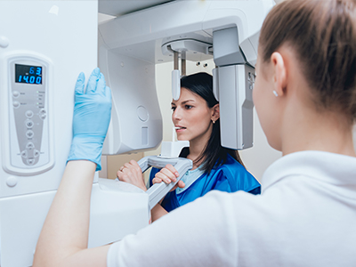
Digital radiography utilizes computer technology and digital sensors for the acquisition, viewing, storage, and sharing of radiographic images. It offers several advantages over the older traditional film based methods of taking x-rays. The most significant of these advantages is that digital radiography reduces a patient’s exposure to radiation. Other benefits are that images can be viewed instantly after being taken, can be seen simultaneously as needed by multiple practitioners, and can be easily shared with other offices. Digital x-rays are also safer for the environment as they do not require any chemicals or paper to develop.
An electronic pad, known as a sensor is used instead of film to acquire a digital image. After the image is taken, it goes directly into the patient’s file on the computer. Once it is stored on the computer, it can be easily viewed on a screen, shared, or printed out.
Digital radiography uses electronic sensors and computer technology to capture dental images instead of traditional film. The sensor records the x-ray image and transfers it immediately to a computer where the image can be viewed, enhanced, and stored. This process streamlines diagnosis and record keeping while eliminating the need for chemical development.
Digital images can be adjusted for contrast, brightness, and detail to help clinicians identify issues earlier and more accurately. Because the images are stored electronically they are easy to organize within a patient record and can be retrieved quickly during follow-up visits. The technology supports safer, faster, and more efficient care for pediatric patients and their families.
The primary difference is the way images are captured and processed: digital systems use sensors and computers while traditional systems use film and chemical development. Digital images appear on-screen instantly and can be manipulated to highlight areas of concern, reducing the need for repeat exposures. Film requires physical storage and chemical processing, which adds time and environmental waste.
Digital systems also enable simultaneous viewing by multiple providers and easier sharing for referrals or specialist consultations. The immediate availability of images improves communication between the dental team and parents by allowing clinicians to show and explain findings in real time. Overall, digital radiography increases clinical efficiency and patient understanding.
When recommended by a pediatric dentist, dental x-rays are considered safe and are taken with strict protective measures in place. Modern digital sensors are more sensitive than film and typically require lower radiation doses to produce a diagnostic image. Clinicians follow the ALARA principle, which stands for "as low as reasonably achievable," to minimize exposure while obtaining clinically necessary information.
Protective tools such as lead aprons or thyroid collars are used when appropriate to shield sensitive areas, and exposure is limited to the smallest number of images required for diagnosis. Routine screening schedules are tailored to each child's risk and development stage so that imaging is used judiciously. Parents are encouraged to ask the dental team about safety protocols and the rationale for any recommended imaging.
Digital sensors are small, flat electronic plates that the clinician positions inside or outside the mouth to capture x-ray images. During imaging the child will be asked to remain still for a brief moment while the x-ray is taken, which usually takes only a few seconds per image. The sensor transmits the image to a computer instantly, allowing the clinician to verify quality and repeat only if absolutely necessary.
Most children tolerate digital imaging well because it is fast and noninvasive, and the team will use child-friendly communication to explain each step. If your child is anxious, the dental staff can use comforting techniques and accommodations to make the visit easier. The overall process is designed to be quick, safe, and as comfortable as possible for young patients.
Digital x-rays provide high-resolution images that reveal details not visible in a standard oral exam, such as cavities between teeth, developing roots, and bone changes. Enhanced image tools allow clinicians to zoom, adjust contrast, and measure anatomical features, improving diagnostic accuracy. Early detection of issues helps guide minimally invasive treatment decisions and can prevent more extensive procedures later.
Because images are available instantly, the dental team can discuss findings with parents during the same visit and create a coordinated plan of care. Digital records also support long-term monitoring of growth and development in pediatric patients. These capabilities make digital radiography a cornerstone of modern preventive and restorative pediatric dentistry.
Digital radiography typically shortens the imaging portion of a dental visit because images are captured and reviewed instantly, eliminating film processing time. Faster image capture reduces the time a child needs to sit still and decreases the likelihood of repeat exposures due to processing errors. The streamlined workflow also helps the overall appointment stay on schedule, which is often appreciated by families.
Comfort is improved by smaller, more ergonomic sensors and the reduced need for positioning adjustments or repeated attempts. The clinical team can focus on gentle behavior management and clear explanations, helping children feel more relaxed. In many cases, the efficiency and speed of digital imaging translate to a calmer, more positive experience for young patients.
Yes, digital images can be securely shared with specialists, referring dentists, or medical providers when necessary and with appropriate patient consent. Electronic sharing eliminates the need for physical film and allows for faster collaboration, which can improve continuity of care. Images are transmitted using secure systems that comply with privacy and health information standards.
Within the dental office, images are stored in the patient record on encrypted or protected systems and are accessible only to authorized staff. Secure storage supports accurate record keeping, follow-up comparisons, and continuity across multiple visits. Parents may request copies or have images sent to another provider as part of coordinated care planning.
Yes, one of the key advantages of digital radiography is that it generally requires less radiation than conventional film to produce a diagnostic image. Digital sensors are more sensitive and efficient, allowing clinicians to capture quality images with lower exposure. This reduction in dose is particularly important when imaging children, who are more sensitive to ionizing radiation.
In addition to lower baseline exposure, careful image selection, proper sensor positioning, and adherence to pediatric imaging guidelines further minimize radiation. The combination of advanced sensors and conservative clinical protocols helps ensure that imaging provides maximum diagnostic benefit with minimal risk.
Yes, pediatric dental teams commonly use intraoral digital sensors for bitewing and periapical images, and extraoral digital systems for panoramic or cephalometric imaging when broader views are needed. In certain cases, three-dimensional imaging such as cone-beam computed tomography (CBCT) may be used for complex diagnostic or surgical planning, but this is reserved for specific clinical indications. Each modality provides different levels of detail and field of view suited to particular diagnostic tasks.
The dental team will recommend the most appropriate type of imaging based on the child’s age, developmental needs, and the clinical question to be answered. Minimizing exposure while obtaining the necessary information remains the guiding principle. Parents should feel comfortable discussing why a particular type of image is recommended and how it will inform treatment.
At Lighthouse Kids Dental we use digital radiography to enhance diagnostic accuracy, streamline visits, and support minimally invasive treatment planning for children. The immediate availability of images allows our team to explain findings to parents and to involve families in shared decision making during the same appointment. Enhanced imaging tools help us detect early signs of decay or developmental concerns so we can recommend preventive measures when they will be most effective.
Digital records also facilitate secure sharing with specialists when interdisciplinary care is required and support long-term monitoring of growth and treatment outcomes. By combining child-focused behavior techniques with advanced imaging technology we aim to provide safe, efficient, and evidence-based care for growing smiles. Parents are encouraged to ask our team about imaging protocols and how results will guide their child’s treatment plan.